The key to revolutionary discoveries in Bacterial Pleomorphism. by Christopher Bird
One day, while waiting for some material to come up from the cellar stacks of the National Library of Medicine in Bethesda Maryland, considerably frustrated by the lack of leads and data concerning the demise of the Rife microscope, I wandered by the Subject card catalogue and casually flipped at random to a card in the middle of a drawer labeled "Microscopes."
The card was filed under "Allied Industries," as if that firm was the author. The company's address was stated to be 4246 Pepper Drive, San Diego, California. The title referenced was "History of the Development of a Successful Treatment for Cancer and Other Virus, Bacteria, and Fungi."2
At the bottom of the card was a single line: "Written by Dr. R. R. Rife."
Entirely by accident, I had stumbled upon what looked to be only one of a series of reports written by Royal Raymond Rife. Fourteen pages long, it was numbered Dev-1042. It was approved and signed by I.F. Crane, manager; Don Tully, development associate; and Verne Thompson, chief electrical engineer.
Are any of these gentlemen alive today?
Was Allied Industries a research corporation established by Rife?
How many other reports did it publish and where are they?
The report so riveted my attention that I was compelled to explore some of the history of microbiology and its connection to cancer and other disease. The present article, much longer than originally planned, is thus the result of a fortuitous finding - perhaps an example of what Jung has called synchronicity - and the consequent preliminary exploration.
Much more needs to be done to tell the story of Rife and his microscope, a fascinating episode in the history of science.
What was the Rife Ultra Microscope?
In February 1944, the Franklin Institute of Philadelphia published an article, "The New Microscopes,"1 in its prestigious journal devoted to applied science.
Founded in 1824 by "philosopher-mechanics," the institute, which recently made studies in its physics laboratory on the best way to move the Liberty Bell to its new Bicentennial Year location, is a smaller analogue of the huge world-famous Smithsonian Institution in Washington, D.C., which reprinted the same article in its own journal shortly after its first appearance.
Authored by R. E. Seidel, M.D., a Philadelphia physician and his research assistant, M. Elizabeth Winter, the essay opened with a six-page discussion of the electron microscope, which had only recently been put on the market by the Radio Corporation of America. This microscope is today standard equipment in modern laboratories.
The article closed with a ten-page treatment of a "Universal Microscope," the brainchild of a San Diego autodidact, Royal Raymond Rife, who developed it with the financial assistance of the rollerbearing and axle magnate, Henry H. Timken, for whose family Rife at one time served as handyman and chauffeur.
Rife's scope, the largest model of which consisted of 5,682 parts and required a large bench to accommodate it, overcame the greatest disadvantage of the electron microscope, its inability to reveal specimens in their natural living state; this is in direct opposition to electron microscopes wherein tiny living organisms put in it are in a vacuum and subject to protoplasmic changes induced by a virtual hailstorm of electrons.
With his invention, Rife was able to look at living organisms. What he saw convinced him that germs could not be the cause, but the result, of disease; that, depending on its state, the body could convert a harmless bacterium into a lethal pathogen, that such pathogens could be instantly killed, each by a specific frequency of light; and that cells, regarded as the irreducible building blocks of living matter, are actually composed of smaller cells, themselves made up of even smaller cells, this process continuing with higher and higher magnification in a sixteen-step, stage-by-stage journey into the micro-beyond.
With the aid of Rife's device, thousands of still pictures and hundreds of feet of movie films were made to reveal these facts, all of this material and the Rife microscopes seem to have disappeared without a trace.
Or have they?
Calls to the U.S. Armed Forces Institute of Pathology Medical Museum, which has hundreds of different microscopes in its historical collection, to the National Library of Medicine's Historical Division, to the Smithsonian Institution and the Franklin Institute (both repositories for outstanding scientific inventions) and to a dozen establishments dealing daily in microscopy elicited from curators, medical pathologists, physicians, and other scientific specialists only the complaint that none of them had ever heard of Royal Raymond Rife and his microscope.
What has become of the Rife microscope? Part 2
The question is not rhetorical. For if even half of the possibilities described for this astounding discovery are true, a massive effort to hunt it down and reactivate its potential might not only save billions of dollars in biological and medical research but open a fascinating new vista onto the nature of life.
From the start, Rife's main goal was to find cures for disease, especially the most intractable of all diseases, cancer. Because he had a hunch that some as yet undiscovered microorganism would prove to play a crucial role in the onset of this malignancy, he tried unsuccessfully to find one by observing all types of malignant tissue with a variety of standard research microscopes.
In the 1920s, it became obvious to Rife that a better means of scrutinizing the microworld than had been developed was indispensable. During that decade, he designed and built five microscopes with a range from 5,000 to 50,000 diameters at a time when the best laboratory microscopes in use could achieve not more than 2,000 diameters of magnification.
At the Rife Research Laboratory on Point Loma, California, he worked at magnifications of 17,000 and higher, to reveal a host of cells and microorganisms never before seen and to photograph them. The work required a saint's patience. It could take the best part of a day to bring a single target specimen into focus.
The Rife microscope had several arresting features: Its entire optical system of fourteen lenses and prisms, as well as an illuminating unit, were made of crystal quartz, which is transparent to ultraviolet radiation. In the scope, light was bent and polarized in such a way that a specimen could be illuminated by extremely narrow parts of the whole spectrum, one part at a time, and even by a single frequency of light.
Rife maintained that he could thus select a specific frequency, or frequencies, of light that coordinated and resonated with a specimen's chemical constituents so that a given specimen would emit its own light of a characteristic and unique color. Specimens could be easily identified, thus solving one of microscopy's greatest (deficiency)...
it was control of illumination that (crossed the necessary barrier to super-efficient observations).
Another feature was the Microscopes extraordinary resolution, its ability to reveal the most minute of component parts of any specimen so that each may be seen distinctly and separately from the others...
Imagine two extremely thin parallel lines. When they can be clearly distinguished, you are still within the microscope's range of resolution.
If the parallel lines blur together, high magnification will only enlarge the distortion and limit of resolution has been attained. With a resolving power of 31,000 diameters - as against 2,000 to 2,500 for the laboratory microscopes in common use in that day, Rife's device could focus clearly on five lines of standardized grid, whereas an ordinary microscope could do no better than examine fifty lines, and that with considerable aberration.
This is somewhat equivalent to one aerial camera's being able to spot individual houses in city blocks from a very great height, while another is able only fuzzily to distinguish the single city blocks themselves.
Controversial Discoveries Beginning in the 1920s and continuing over seven years, Rife and his colleagues worked on more than 20,000 laboratory cultures of cancer obtained from the Paradise Valley Sanitarium in National City, California, in what appeared at first to be a fruitless effort to isolate microorganisms that he felt should somehow be associated with the disease.
Up until then, bacteria had clearly been proven to be linked with a wide variety of ills including tuberculosis, leprosy, cholera, gonorrhea, syphilis, typhoid, bubonic plague, pneumonia, and others. But no one had found them in association with cancer.
In contrast to the much smaller viruses, bacteria were widely considered to be unicellular, monomorphic (meaning one shape and one shape only) forms. A quarter of a million of them can occupy a space no larger than the period at the end of this sentence. They come in various shapes. Cocei are round, bacilli rod-like, to offer two examples:
There are various forms for each shape. Of the round-shaped ones, monococci appear singly, diplococci come in pairs, staphylococci in clusters resembling a bunch of grapes, streptococci, which under certain conditions can produce a painful sore throat, in chains.
While outside a host, or body, bacteria are hard to raise, or culture. Each type has been studied as a pure culture type by isolating it upon a specific nutrient called media.
Bacteria also have specific maximum, minimum, and optimum temperatures in which they will live and multiply. Some, like polar bears, are addicted to arctic temperatures and even live in ice. Others prefer water so hot it would kill most animals (i.e. extremophiles - let's not forget the bacteria living in the vaccuum of space, the asteroid belt of our solar system). A great many enjoy the temperature of the human body. Millions of them are living, harmlessly, inside you right now...
But they are not always harmless. They can acquire virulence, or the power to cause disease, under some conditions but not others, although even today no one knows exactly why.
This mystery, in the 1920s, was closely connected to a debate in microbiology so hot as to seem almost a war. On one side were those who affirmed - as do many textbooks today - that bacteria were eternally monomorphic. They could not assume other or smaller forms, as small, say, as a virus.
Originally, virus - the word means "poison" in Latin - was the name generally applied to any microscopic agent injurious to living cells. Now it is much more narrowly defined as "one of a unique group of very small infectious agents that - grow only in cells of animals (including humans), plants, and also bacteria."
Because they were so small, viruses would pass through filters that did not allow the passage of bacteria, said to be monomorphic, just as a net of small enough mesh will allow minnows to pass through it but bring the fish that are preying upon them up short. It is this filter-passing ability of viruses that is widely held today - along with their inability to grow on artificial media - to be one of the main criteria separating them from bacteria.
For several decades, however, another school of microbiologists maintained that, far from holding everlastingly to one shape, bacteria were pleomorphic, or form changing. They could be caused, under the right conditions of culture, to metamorphose into forms small enough to pass through filters just like viruses.
Because of their sharp disagreement on the filterability of bacteria, the two camps came to be called "filtrationist" and "nonfiltrationist."
One of the earliest of the filtrationists was a Swedish physician and explorer, Ernst Bernhard Almquist,10 for whom islands off the north Siberian coast are named. Almquist made hundreds of observations of pleomorphic bacteria in his laboratory as did researchers in Italy, Russia, France, Germany, and the United States. In 1922, after two decades of work, Almquist came to the conclusion that "nobody can pretend to know the complete life cycle and all the varieties of even a single bacterial species. It would be an assumption to think so."
Way back in 1914, the American bacteriologist Dr. Edward C. Rosenow3,4 had the gall to assert (to conventional scientists) that bacteria were not unalterable and that various strains, or what one might call sub-subspecies of them, could, when suitably treated, become any of the other strains. It was Rosenow's contention, too, that he found a form of the streptococcus bacterium which caused poliomyelitis, commonly known as infantile paralysis.
What Rife's opinions were about this heated controversy are not known. He followed the standard bacteriological practice of the day, first implanting small patches of cancer tissues on various nutritive media including a special "K" medium developed by another filtrationist, Dr. Arthur Isaac Kendall,8,9 at the Northwestern University School of Medicine in Chicago, Illinois. The medium, which bore the first letter of Kendall's name, seemed to have the faculty of transforming bacteria into the transitional forms alleged for them by the filtrationist school, No matter how often he changed menus for his sought-after cancer microbe, no matter how he altered the temperature of incubation, Rife seemed unable to coax it to appear in his cultures.
Rife discovered that many microbes respond to the effects of light from noble gases, such as neon, xenon, and argon, by changing their growth patterns...
It was apparent only when, as a result of his continuing physical experimentation with the effects of light frequencies, that Rife hit upon a solution to the problem that was nagging him; he discovered that many microbes respond to the effects of light from noble gases, such as neon, xenon, and argon, by changing their growth patterns.
He placed a sealed test tube containing cancer tissue into a closed loop filled with argon gas. After creating a vacuum within the loop, he charged the gas with electricity, just as one does when one throws the switch to light up the neon lamps in modern offices. In Rife's case the charge was 5,000 volts. While he still could not reveal any microbes, he noted a certain cloudiness in the nutritive medium, which, through chemical analysis, he ascribed to ionization caused by the electronic bombardment.
Readers may well wonder why he adopted so strange and novel a process. The question is just as unanswerable as if put about Rife's next step: In order, he said, to counter the ionization, he placed the tube into a two-inch water vacuum and heated it for twenty-four hours at near body temperature.
(In this webmaster's humble opinion, Rife was reproducing the bioenergetic forces of the living body within the medium. Imbalanced changes in bio-electrical potential within the body are a precursor to disease; this concept applies to bioresonant energy medicine such as photonics and bioenergetic biofeedback which induce balanced patterns towards homeostasis.)
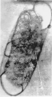
Under his microscope, at 20,000 X, the tube now teemed with animated forms measuring only 1/20 by 1/15 of a micron, much smaller than any known bacteria. They refracted a purplish red color in the specific (frequency of) light beam.
He called this form Bacillus X and, later, because it was so much smaller than other bacilli, and perhaps because of the filterability controversy, BX virus. This problem of nomenclature can be resolved herein by referring to Rife's organism as a BX form, or simply BX.
Rife writes that "this method of ionization and oxidation brought the chemical refraction of BX out of the ultraviolet and into the visible band of the spectrum. Owing to the fact that the test-tube specimens had gone through so many trials, we again started from scratch and repeated this method 104 consecutive times with identical results."
Because he could culture his BX form small enough to pass through any filter, he seemed to have discovered a filterable form of a bacterium... [BX] could change into not just fairly similar forms as Rosenow had previously discovered, but into completely different forms simply by altering the medium on which they were living only very slightly... (a direct explanation for specific cancer biopathies according to individual constitution!)
But just finding bacteria, even in filterable form, in a human tumor does not necessarily imply that they are its cause. To make sure, it is held they must be reinjected into animals and seen to cause the same or nearly similar disease, after which they must then be reisolated and shown to resemble the original organism. These were the postulates propounded by the German pioneer bacteriologist, Robert Koch, who proved that tuberculosis was apparently caused by the tubercule bacillus.
Following this accepted procedure, Rife inoculated the new BX forms into over 400 rats in all of which there subsequently appeared "tumors with all the true pathology of neoplastic tissue."
Some of the tumors became so large they exceeded the total weight of the individual rats in which they were developing. When the tumors were surgically removed, the BX form was recovered from them in all cases. Koch's postulates were fulfilled.
More Startling Discoveries: How the host morphs bacteria into other life forms and how these life forms are affected by monochromatic light.
By continued microscopical study and repeated photography to stop their motion, Rife and his co-workers next came to the baffling conclusion that the BX, far from remaining always what he had seen as the purplish red bodies a fraction of a micron in dimension, could change into not just fairly similar forms as Rosenow had previously discovered, but into completely different forms simply by altering the medium on which they were living only very slightly.
"Slightly" in Rife's case meant an alteration in the nutrient environment of only two parts per million (ppm) by volume. Those who would consider this unlikely may recall that in homeopathic medicine doses of remedies are given in dilutions of this weakness and beyond. Even though they have nothing chemically analyzable in them, they are effective. (webmaster note: Another consideration is the sensitivity of the integral bioenergetic response to the slight chemical alteration in terms of morphic fields using mitogenic radiation.)
One such alteration caused the BX to become what Rife called a Bacillus Y, or BY. It was still the same purplish red color as the BX but so enlarged that it would not pass through a filter.
With the second change of the medium, the BY enlarged still further into a monococcoid or single disk form which, when properly stained, could be viewed under a standard research microscope. Rife claimed that these forms could be found in the blood of over ninety percent of cancer victims.
By removing this form from the fluid medium it inhabited and depositing it onto a hard base of asparagus or tomato agar, Rife then saw it miraculously develop into a fungus, making it kin to a yeast, mold, or mushroom. (webmaster note: This observation would verify the morphic field integral response within organisms according to environment.)
Any of these succeeding forms, Rife stated, could be changed back within thirty-six hours into a BX form capable of producing cancer tumors in experimental animals from which, in turn, the same BX form could again be recovered.
The transformation did not stop with the fungus, which, if allowed to stand dormantly as a stock culture for a year and then replanted onto the asparagus medium, would then change into bacillus coli, millions of which live in the human intestine. This common bacillus could pass, in Rife's words, "any known laboratory method of analysis."
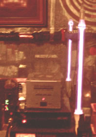
Because he had found that microorganisms had the ability to luminate when stimulated by given frequencies of light, it occurred to Rife that they might also be devitalized by beaming radiations of specific frequencies upon them...
One source (?) has it that the harmonics of these frequencies ranged from 10 meters to 20,000 meters (in wavelengths, i.e. speed of light = wavelength/frequency; frequency = wavelength/speed of light; ƒ = ƚ/c).
To this end, he had been developing concurrently with his microscopic equipment a special frequency emitter, which he continued to improve, up to at least 1953, as steady advances in electronics continued. The killing waves were projected through a tube filled with helium gas and said to be efficient in destroying microorganisms at a distance of as much as one thousand feet.
With this device, he noted that when the proper mortal oscillatory rate (MOR) was reached, many lethal organisms such as those of tuberculosis, typhoid, leprosy, hoof-and-mouth disease, and others appeared to disintegrate or "blow up" in the field of his microscope. This "death ray" principle was also effective when applied to cultured BX.
The obvious next step was to determine whether similar radiation would affect the BX, not in culture, but in the bodies of cancer-afflicted animals. It apparently did so, for Rife states he got rid of BX in over 400 experimental rats and other animals in his lab. If it worked on animal cancers, wondered Rife, why not on human cancers?...
The answer was so resoundingly "Yes" that, in our day when billions are being spent each year to find a cure for cancer, it is prudent to quote Rife's report word-for-word:
The first clinical work on cancer was completed under the supervision of Milbank Johnson, M.D., which was set up under a special medical research committee of the University of Southern California. Sixteen cases were treated at the clinic for many types of malignancy. After three months, fourteen of these so-called hopeless cases were signed off as clinically cured by a staff of five medical doctors and Alvin G. Foord, M.D., pathologist for the group. The treatments consisted of three minutes duration, using the frequency instrument which was set on the mortal oscillatory rate for BX, or cancer, at three-day intervals. It was found that the elapsed time between treatments attains better results than cases treated daily.
News of Rife's work began to leak out to the world of medicine at the end of the 1920s. One of the first to learn of it was Arthur W. Yale, M.D.,5 who lived in San Diego, not far from Rife's laboratory. He acquired a frequency emitter and began to treat cancerous patients.
In 1940, reporting to his fellow physicians on some of his decade-long results, Yale wrote that because the whole of Rife's extraordinary findings constituted an "entirely new theory of the origin and cause of cancer, and the treatment and results have been so unique and unbelievable," he was making his findings available in the hope that "after further research we may eliminate the second largest cause of deaths in the United States."
(webmaster note: The U.S. Gov't had a completely different agenda in the 1940's when certain Nazi scientists where imported to expand their experiments in artificial fluoridation on the public water system.)
Yale had had limited success in treating cancerous tumors with X rays and with the use of what he called "static wave current" for some three decades. When he began to use Rife's device, he sometimes employed it alone, sometimes together, with the two methods with which he was familiar. Both methods brought startlingly successful results. Yale was careful to note that, when he added the use of the Rife ray to his other radiation, cancerous masses "have disappeared in about one-tenth the time and so far with no reoccurrences."
Dr. Arthur Isaac Kendall, whose "K" medium Rife had used in his experimentation, was also determined to check whether viable bacteria in the filterable state could be unequivocally seen by Rife's microscope. Kendall had been working with cultures of typhoid bacillus and, under a standard microscope, had been able to detect a swarm of active granules that could be seen only as tiny motile points. Because nothing of their individual structure could be ascertained, Kendall could not diagnose them with certainty to be filterable forms of the bacillus.
In order to make certain, he went to California in late November of 1931 and examined his cultures under a Rife microscope at 5,000 diameters in the Pathological Laboratory of the Pasadena Hospital. The facilities were afforded through the offices of the same Drs. Johnson and Foord who had worked with Rife on the BX.
When Rife finally got them in focus, the tiny granules were seen to be bright, highly motile, turquoise-blue bodies, which, to quote the report he coauthored with Kendall, "contrasted strikingly both in color and in their active motion with the noncolored debris of the medium." The same observations were repeated eight separate times, the complete absence of similar bodies in uninoculated control media being noted.
To further confirm their findings, Rife and Kendall next examined eighteen-hour-old specially cultured and inoculated colonies of the same bacillus because they had determined that it was precisely at this stage of growth that they became filterable. Now they could see three transitional forms of the same organism: one, the normal bacillus itself, almost devoid of color; two, the same bacillus but with a prominent turquoise blue granule at one end of it; and three, the same turquoise blue granules moving about independently.
This was somewhat equivalent to being able to observe a caterpillar, its cocoon, and the butterfly that emerges from the cocoon, all simultaneously.
When they transplanted the filter-passing granules into a broth medium, they were seen under the, Rife microscope to revert back to their original bacillus, or rod-like, form.
At this juncture, the American bellwether journal Science got wind of Kendall's work and, in a news story devoted to it, referred to the new "supermicroscope" invented by Royal Raymond Rife.
The same month, December 1931, the Rife-Kendall account was published in California and Western Medicine, the official mouthpiece of the state medical associations of California, Nevada, and Utah. This magazine also commented editorially that the Kendall-Rife article was to be particularly recommended to its readers because of its "calling the attention of the world to a new type of microscope which, if it fulfills its apparent advantages over any microscope thus far developed, bids fair to lay the basis for revolutionary discoveries in bacteriology and the allied sciences."
The editorial was significantly entitled "Is a New Field About to Be Opened in the Science of Bacteriology?" Apparently it was about to die aborning.
The Opposition Mounts: Scientific Cretinism + Anger in the radical left mainstream
The following month, Kendall was invited to give the De Lamar lecture at the Johns Hopkins University School of Hygiene and Public Health in Baltimore, Maryland, before the Association of American Physicians. As a leader of the filtrationist school, he attracted the attention of his adversaries, two of whom were invited as discussants.
The first was an irascible, pugnacious curmudgeon, Dr. Thomas Rivers, of the well-heeled Rockefeller Institute of New York City, who was described by one of his institute colleagues as a "difficult and formidable person to oppose and [he] could be stubbornly inflexible in maintaining a position."
When he learned of his invitation to discuss Kendall's presentation of the work with the typhoid bacillus, Rivers hurriedly repeated experiments on which Kendall had worked for years and, by his own account, got no proof of Kendall's claim. Based on this thin evidence, he arose at the Johns Hopkins meeting and, to quote him "in a very temperate manner called the fellow a liar. Not in so many words. Actually, all I said was that I couldn't repeat this experiment and I therefore didn't believe his findings were true."
Rivers was followed in the discussion by the Harvard microbiologist, Dr. Hans Zinsser, also a "nonfiltrationist," who, to quote Rivers anew, "just gave Kendall bloody hell. I'd never seen Hans so hot in my life. I had to agree with everything he said - but I really felt sorry for poor old Kendall he just sat there and took it."
In the midst of the venom and acerbity, the only colleague to come to Kendall's aid was the grand old man of bacteriology, and first teacher of the subject in the United States, Dr. William H. "Popsy" Welch, who evidently looked upon Kendall's work with some regard.
..it was curious that Rivers could claim to have repeated Kendall's work without the use of the instrument (Rife Microscope) Kendall had found so necessary to clearly reveal his filterable forms... (Obviously, Rivers did not repeat Kendall's work.)
What is of interest today is that at the Baltimore meeting there seemed to be no mention of the Rife microscope. Also, in the light of the apparent victory of the "nonfiltrationists" over those who claimed that bacteria were filterable, it was curious that Rivers could claim to have repeated Kendall's work without the use of the instrument Kendall had found so necessary to clearly reveal his filterable forms.
Kendall's work, however, attracted the rapt attention of the same Dr. Edward C. Rosenow3,4 who, in 1914, had been able to prove that strains of streptococcus were able, under the right conditions, to transmute one into the other. In that day, he had written that these "conditions were more or less obscure. They seem to call forth new or latent energies which were previously not manifest and which now have gained the ascendancy."
As a filtrationist, Rosenow was a maverick among bacteriologists up to his death at ninety-four in the 1960s. His work had convinced him, also prior to World War I, that organisms in sera - the fluids from tissues of immunized animals commonly used as antitoxins to neutralize microbes in the body - might in some patients have dangerous biological side effects.
The main implication of Rosenow's work in his own eyes was that bacteria were not as important to disease as the terrain on which they found themselves. "It would seem," he wrote in his 1914 article, "that focal infections are no longer to be looked upon merely as a place of entrance of bacteria but as a place where conditions are favorable for them to acquire the properties which give them a wide range of affinities for various structures."
Rosenow first became aware of the Rife technique through a patient at the Mayo Clinic in Rochester, Minnesota, where Rosenow was employed. The patient was none other than the same Henry H. Timken, who had financially aided Rife to develop his microscope and begin his research in the 1920s.
"The oval, motile, turquoise blue bodies," wrote Rosenow of this work, "described previously by Kendall and Rife were unmistakably demonstrated..."
Rife came to Chicago with his microscope. Kendall invited Rosenow down to the Northwestern University Medical School to work with himself and Rife on 5 May 1932. For three days, they made a restudy of the Kendall forms, Rosenow working with a Zeiss microscope, Kendall with an oil immersion dark-field instrument, and Rife with his special device. "The oval, motile, turquoise blue bodies," wrote Rosenow of this work, "described previously by Kendall and Rife were unmistakably demonstrated."
The three next decided to filter cultures of the streptococcus bacteria that Rosenow had found to be associated with poliomyelitis to see what the Rife scope might reveal. What they saw were not the blue bodies linked to the typhoid bacillus, but cocci and diplococci of a brownish gray color each surrounded by a strange halo. These could only be observed in the Rife microscope.
Moreover, filtrates of a virus considered to be the cause of encephalitis showed a considerable number of round forms, singly and in pairs, which under the special Rife illumination were pale pink in color and somewhat smaller than those seen in the poliomyelitis preparations.
Rosenow's work was panned by Rivers in public forum just as viciously as was Kendall's. This was before Rosenow had worked with the Rife microscope. "I had one run-in with him," said Rivers, "at a meeting held before the Association for Research in Nervous and Mental Diseases during Christmas week, in 1931. I was pretty savage with him. Do you think that helped? Hell, no, if you ask me for my candid opinion, I think that most of the audience believed Rosenow."
This belief did not last for long. For a variety of reasons, including the very difficult methods of culturing the filterable forms of bacteria - and lack of the Rife microscope to observe them - the "church" of nonfiltrationist bacteriology, of which Rivers was later proclaimed "the apostolic father" (does one need better evidence of hierarchical priesthoods and priestcraft in science?), was putting the filtrationist camp on the defensive.
Three filtrationists, writing of discoveries similar to those of Kendall, just prior to Kendall's Johns Hopkins lecture, thus considered it necessary to state in their introduction: "It has come about these days that to express convictions that differ from the consensus gentium becomes almost professional foolhardiness: It brings down the strictures of one's friends and enemies alike."
They added: "But we are also conscious of the fact that, beneath the tumult of controversy between monomorphism and pleomorphism, there is being born a new epoch in bacteriology, the limits of the significance of which and the possible expansion of which no one can yet surmise."
Like all scientific revolutions, the epoch would have to wait patiently for its time to come. Rosenow was held by his adversaries to be 100 percent wrong in many of his observations. His son, Dr. Edward C. Rosenow, Jr., chief administrative officer of the American College of Physicians, asserts that his father was all but accused by Rockefeller Institute research moguls of experimental dishonesty.
How was it that none of Kendall's or Rosenow's attackers bothered to use the Rife microscope? Rife himself admitted that he was not confident that his experiments, revealing the BX form, could ever be repeated without the use of his scope. "We do not expect any laboratory," he wrote, "to be able to produce the BX on account of the technique involved and adequate optical equipment. This is why we have never publicly announced that BX is the cause of cancer but we have succeeded in producing from its inoculation tumors with all the true characteristics and pathology of neoplastic tissue from which we have repeatedly recovered the BX virus."
At the end of his life, Rosenow was philosophic about lack of acceptance for his findings among his colleagues. "There is no way," he told his son, "to convince one's peer group of something new until their attitude of receptivity changes. They simply won't listen." This echoes the German Nobel Laureate in physics Max Planck, who stated that for new ideas to be accepted, one had to wait for a generation of scientists to die off and a new one to replace it. (Unfortunately, the generation of which Planck speaks has already trained a "new" generation to think the same way...)
The Search Continues: The "rediscovery" of pleomorphism
With respect to Rife's cancer observations, it may be that this process of replacement is now taking place.
Rife's work has a possible connection with research performed over the last twenty years by several pioneers. One pair of them are Dr. Irene Diller,20 a former long-time associate of the Institute for Cancer Research in Philadelphia, and Dr. Florence B. Seibert,13,21,22 professor emeritus of biochemistry, University of Pennsylvania.
One day in the late 1950s, Diller called Seibert, who won many awards and five honorary doctorates for her more than thirty-year-long work on tuberculosis, and asked her to come and look at some microbes on slides. On the slides, Seibert observed tiny round organisms. When Seibert learned that Diller had isolated them regularly from many other tumors, as well as from the blood of leukemia patients, she hastened to ask whether Diller could find them in a sarcoma tumor she, Seibert, was studying.
After several weeks, Diller showed Seibert a tube filled with a slightly grayish and moist-looking culture fined with small round cocci. Injected into mice, they produced cancerous tumors.
Seibert became convinced that Diller might have found a link to cancer. Because so many scientists, believing Diller's new forms to be merely "ubiquitous contaminants" in her cultures, were writing off her work as spurious, Seibert decided to continue working on the problem during her Florida retirement, first at the Mound Park - today the Bay Front - Hospital in Saint Petersburg, later at a Veterans Administration Hospital.
Blood samples from cancer patients with varying types of leukemia were obtained and from every one of them Seibert was able to isolate pleomorphic microbes. These bacterial forms were also isolated from tumors, and with a homologous vaccine they decreased tumors in mice.
Just like those of the Rife-Kendall-Rosenow research, they could change from round to rod shaped and even could become long threadlike filaments, depending on what medium they were grown in and for how long. They would pass a filter and at this stage in their life cycle they were about the same size as Rife's BX forms.
Today there is great stir about, and much money devoted to, viruses in relation to the cancer problem. The most recent edition of the Encyclopedia Britannica states that "sufficient evidence has been acquired to indicate that one or more viruses probably cause cancer in man," and that carcinogens, or cancer-producing agents, "are suspected of producing cancers by activating viruses latent in the body."
But, so far, little support is given to those who ascribe bacteria and the forms into which they transmute the ability for close association with cancer. This legacy of the nonfiltrationist school persists in the face of mounting evidence that the filtrationists may have been right all along.
These days, because various bacterial forms have been noted to have anomalies in their cellular walls - how could they develop into smaller forms if they could not leap beyond or through the walls that imprison them?
They are known as Cell Wall Deficient Forms. A revolutionary new book about them has been written by the Wayne State University microbiologist Dr. Lida H. Mattman,14 Her text opens with the statement: "Clandestine, almost unrecognizable, polymorphous bacterial growth seems to occur as often as the stereotyped classical boxcars of bacilli and pearls of cocci ..." The book's contents would seem to indicate that the new era predicted in 1931 for filtrationist microbiology is dawning, though presently its adherents are having great difficulty both in publishing their work and getting grants for further research.
Sufficient data, writes Mattman, have been amassed to warrant reinvestigation, and adds: "There is no subject generally viewed with greater skepticism than an association between bacteria and human cancer. However, the medical profession may look back with irony at the stony reception given by his home colleagues to Koch's paper elucidating the etiology of tuberculosis. Similarly, medical students were once taught that whooping cough vaccination was an unrealistic dream reported only by two women at the Michigan Public Health Laboratories and by a pediatrician namer Sauer."
Most importantly, she concludes: "One must always consider that most malignancies are accompanied by an immunodeficiency ... Therefore, we could be dealing with a microbe that finds such a host merely a suitable environment for habitation."
This is very close to Rife's own statement that he had unequivocally demonstrated that "it was the chemical constituents and chemical radicals of an organism which enacted upon the unbalanced cell metabolism of the human body to produce disease." Before he died, Rife stated: "We have in many instances produced all the symptoms of a disease chemically in experimental animals without the inoculation of any virus or bacteria into their tissues."
What of Royal Raymond Rife and his microscope? Part 3
Lingering Questions on the Origins and Properties of Cancer:
How is it that biologists and physicians, other than Kendall and Rosenow, did not rush to investigate it? Why haven't (bio)physicists looked into the effects Rife achieved with electromagnetic waves of specific frequencies upon disease, including cancer?
Similar effects were observed by Dr. Georges Lakhovsky16 in Paris, who developed a wave emitter called a multiwave oscillator with which he cured cancer as well as other diseases in plants and humans. The multiwave oscillator is today banned by the FDA as quackery.
They have also been noted in Bordeaux by another inventor, self-taught as was Rife, Antoine Priore, whose apparatus combines the use of electromagnetic radiation with a plasma of helium or noble gases reminiscent of Rife's method used in detecting and devitalizing BX.
Are the strange blue, motile forms that Dr, Wilhelm Reich17 discovered in the late 1930s and for which he coined the word, "bions" related to the foregoing? Reich observed the bions to spontaneously proliferate from specially treated organic matter and even from inert matter (coal and sand!) Spontaneous generation of life was supposed to have been laid to rest in Reich's time, as it is in ours, and he was accused by fellow scientists of confusing Brownian movement of subcellular particles or debris in his cultures with the new subcellular forms he claimed to have discovered.
In cancerous patients, Reich observed the bions to degenerate into what he called T-bacilli (the T coming from the German word Tod, meaning death). When injected into mice, they caused cancer just like Rife's BX forms.
In Copenhagen, a biophysicist named Scott Hill reports that a new book written in Russian by two researchers at the Kazakh State University in the U.S.S.R. deals with a whole new branch of medical science in which "healing" of various disorders is being accomplished by the use of ultraweak, monochromatic laser light. (Shades of Rife! See paper on low level laser therapy.)
The Lee Foundation for Nutritional Research in Milwaukee, Wisconsin maintains that Rife, his microscope, and his life work were tabooed by leaders in the U.S. medical profession and that any medical doctor who made use of his practical discoveries was stripped of his privileges as a member of the local medical society.
The assistance of NAJ readers is solicited. After the above article was published, further investigation located Rife's "Universal Microscope" in a sorry state of disrepair in the San Diego home of John Crane. Efforts to rebuild it have so far been unsuccessful. A fascinating book on Rife's saga, The Cancer Cure That Worked, by Barry Lynes, was published in 1987 by Marcus Books, Toronto, Canada.
References:
Seidel, R. E., and M. Elizabeth Winter. "The New Microscopes," Journal of the Franklin Institute, February 1944.
Allied Industries, "History of the Development of a Successful Treatment for Cancer and Other Virus, Bacteria and Fungi," Report no. DEV-1042, 1 December 1953, written by Dr. R. R. Rife.
Rosenow, E. C. "Transmutations Within the Streptococcus-Pneumococcus Group," Journal of Infectious Diseases, vol. 14, 1914.
Rosenow, E. C. "Observations on Filter-Passing Forms of Eberthella Typhi (Bacillus Typhosus) and of the Streptococcus From Poliomyelitis," Proceedings of the Staff Meetings of the Mayo Clinic, 13 July 1932.
Yale, Arthur W. "Cancer," Pacific Coast Journal of Homoecopathy, July 1940.
"Filterable Bodies Seen With the Rife Microscope," Science Supplement, Science, 11 December 1931.
"Is a New Field About to Be Opened in the Science of Bacteriology?" Editorial, California and Western Medicine, December 1931.
Kendall, Arthur Isaac, and Royal Raymond Rife. "Observations on Bacillus Typhosus in its Filterable State," California and Western Medicine, December 1931.
Kendall, Arthur Isaac. "The Filtration of Bacteria," Science, 18 March 1932.
Almquist, E. Biologische Forshungen Weber die Bakterien (Biological Research on Bacteria), Stockholm, 1925.
Benison, Saul, and Tom Rivers. "Reflections on a Life in Medicine and Science," an oral history memoir prepared by MIT Press, 1967.
Hadley, Philip, Edna Dalves, and John Klimel. "The Filterable Forms of Bacteria," Journal of Infectious Diseases,. vol. 48, 1931.
Seibert, Florence B. Pebbles on the Hill of a Scientist, self-published, Saint Petersburg, Florida, 1968.
Greenberg, Daniel S. "The French Concoction," Esquire, July 1975 (full account of Antoine Price and his invention).
Lakhovsky, Georges. La Formation Neoplastique et le Desequilibre 0scillatoire Cellulaire (Neoplastic Formation and Cellular oscillatory Disequilibrium). Paris: G. Doin, 1932.
Reich, Wilhelm. The Cancer Biopathy. New York: Orgone Press, 1948.
"The Rife Microscope of Facts and Their Fats," Reprint no. 47, The Lee Foundation for Nutritional Research, Milwaukee, Wisconsin.
Inyushin, V. M., and P. R. Chakorov. Biostimulation Through Laser Radiation and Bioplasma, Kazakh State University, U.S.S.R. (in Russian).
Diller, Irene, "Tumor Incidence in ICR-Albino and C37/B16JNicr Male Mice Injected With Cultured Forms From Mouse Malignant Tissues," Growth, vol. 38, 1974, page 507.
Seibert, F. B., F. M. Feldmann, R. L. Davis, and I. S. Richmond, "Morphological, Biological, and Immunological Studies on Isolates From Tumors and Leukemic Bloods," Annals of the New York Academy of Sciences, vol. 174, 1970.
Seibert, F. B., "Decrease in Spontaneous Tumors by Vaccinating C3H Mice With an Homologous Bacterial Vaccine," International Research Communications Service, vol. 1, 1973.
(PhysOrg.com, October 14, 2010) -- Cancer is a modern, man-made disease caused by environmental factors such as pollution and diet, a study by University of Manchester scientists has strongly suggested.
Nikola Tesla's Magnifying Transmitter Massively Increased It's Electrical Power - Tesla Even Did This With Ordinary Generators... But Never Revealed The Secret. Finally, The Code Has Been Cracked And A Working Model Has Been Demonstrated That Multiplies The Input Power By 4790%!!! [May 2020 Shelter in Place Special - EXPIRES MAY 31st: Get 50% off all 2018-19 A&P Presentations, using coupon code, MAY50 (case sensitive), in the shopping cart for each purchase.] .
Water Nanobubbles Are Surprisingly Stable; They Become Templates For Creating Microscopic Ball Lightning In A Combustion Chamber. This Yields The Simplest "Excess Energy" Machine For Hobbyists.
The Tesla Transmitters are designed to replicate the ideas expressed in Wilhelm Reich's "Cosmic Superimposition" of formative fields. When two living energy fields mutually interact, they can stimulate each others' excitation to the point of illumination - in other words, CREATE LIGHT!


Comments